
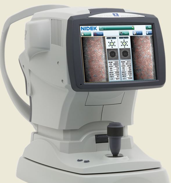
Nidek CEM-530 Specular Microscope
Original price was: $4.730.$2.690Current price is: $2.690.
All three manual analysis methods can be performed on the same image and also on auto-analyzed images. The versatility of combining automated and manual analyses allows analysis of the range of pathology in a comprehensive practice.
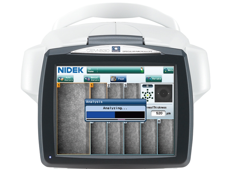
The CEM-530 with new advanced software and enhanced image capture system allows rapid image acquisition.
Description
Nidek CEM-530 Specular Microscope
The analysis results with graphic and color-coded cell images helps the clinician to rapidly and effectively evaluate the endothelial cell layer.
In addition to conventional central and peripheral specular microscopy, the CEM-530 includes a unique function to capture paracentral images.
- Paracentral specular microscopy
- Faster measurements and two-second auto analysis
- Comprehensive analysis
- Advanced manual analysis functions
- Easy operation
- Additional features with CEM Viewer for NAVIS-EX
Combination of auto and manual analyses
All three manual analysis methods can be performed on the same image and also on auto-analyzed images. The versatility of combining automated and manual analyses allows analysis of the range of pathology in a comprehensive practice.
Features
- Multi area specular microscopy
- Enhanced usability and quick analysis
- Advanced manual analysis functions
- Combination of auto and manual analyses
- Additional features with CEM Viewer for NAVIS-EX
Multi area specular microscopy
In addition to conventional central and peripheral specular microscopy, the CEM-530 includes a NIDEK original function that captures paracentral images. The combination of central, paracentral, and peripheral imaging provides a broader, overall view that can be used for detailed morphological and quantitative evaluation of the endothelial layer and individual cells.
Enhanced usability and quick analysis
The 3D auto tracking and auto shot functions result in a user friendly and patient friendly experience.
Data analysis within 2 seconds allows efficient patient flow.
Advanced manual analysis functions
Center point
Select the approximate center of a cell. The cells are detected based on the surrounding points. This method is effective for areas where groups of cells are clumped together.
Corner point
Trace the outlines of the cells to be analyzed by selecting the corners of each cell. This method is suitable for detailed identification of the size and dimension of isolated cells.
Pattern select
Select a hexagonal reference pattern that is similar to the cell size and drag it onto the cell to be analyzed. This method is effective for rough identification of the size and dimension of the cells.
Combination of auto and manual analyses
All three manual analysis methods can be performed on the same image and on auto-analyzed images. The versatility of combining automated and manual analysis on the same image allows for better clinical interpretation of the diverse range of pathology in a comprehensive practice.
dditional features with CEM Viewer for NAVIS-EX* (optional)
CEM Viewer is software used for viewing and working with CEM-530 data via NAVIS-EX. This function enhances the capability of the CEM-530 with additional features and increases the efficiency of any clinic.
*NAVIS-EX is optional software and is required for use of the CEM Viewer.
Data management and endothelial cell count
Unlimited NAVIS-EX database is available for review on the CEM Viewer. The basic functions of the CEM-530 such as endothelial cell count are available on the CEM Viewer.
Progression follow-up and comparison
Multiple examination data sets are displayed in chronological order for follow-up. Additionally, two data sets are displayed for comparison. Endothelial changes can be monitored over time with this function.
Paracentral display with peripheral
The images and analyses of the paracentral and peripheral areas are displayed providing a comprehensive image of endothelial cells.
Related products
-
- Sale!
- : Beauty Equipment, facial care, Laser & Aesthetics, Laser Equipment, Medical Equipment, Medical Laser, Medical Products
2019 Syneron Candela Profound Radio Frequency Microneedling
- Original price was: $11.310.$5.104Current price is: $5.104.
- Add to cart
-
- Sale!
- Body Contouring, Medical Equipment, Medical Products, Surgical
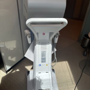
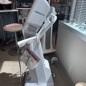
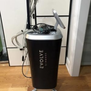
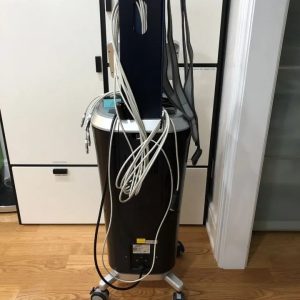
Inmode Aesthetics Evolve
- Original price was: $8.699.$5.270Current price is: $5.270.
- Add to cart
-
- Sale!
- Beauty Equipment, Medical Products, Skin Rejuvenation
IDS Daeshin Shiny
- Original price was: $5.145.$2.810Current price is: $2.810.
- Add to cart


Reviews
There are no reviews yet.